Uncharted Warming
The earth has just witnessed its hottest January on record, defying expectations and leaving climate scientists scrambling for answers.

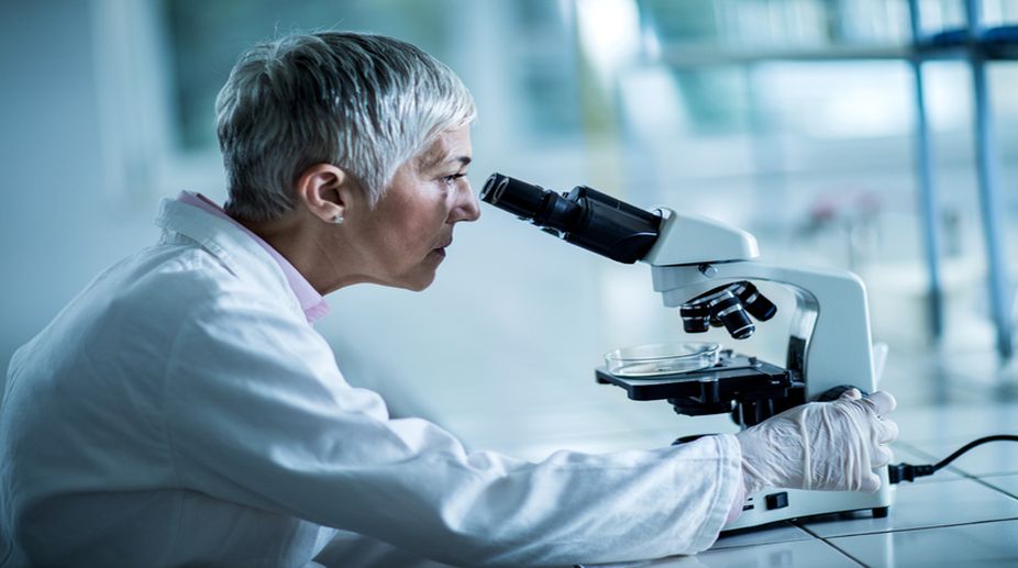
Optical Microscopes (PHOTO: GETTY IMAGES)
Early scientists used an item that was destined to be omnipresent in a laboratory — the microscope. 70-yearold Bradshaw Amos has spent most of his time studying a different and very tiny world, also through a microscope. He is a visiting professor, at the University of Strathclyde, Scotland, and works with a team of researchers; they are in the process of designing a large, new, microscope lens, about the length and width of a human arm and the lens is perceived as a significant innovation. The Mesolens is so powerful that it can image entire tumours or even mouse embryos, in one field of view, while simultaneously imaging the insides of cells.
For hundreds of years, optical microscopes have allowed living tissues to be studied in fine detail. Unfortunately, images captured through a typical microscope lens feature a compromise between the level of detail in the image and how much of the sample can be shown. Densely packed individual cells cannot be distinguished in an image that shows an entire mouse embryo. This accentuates the importance of the Mesolens because it can magnify samples to four times, in higher detail than conventional lenses.
Advertisement
Current optical microscopes, which have a low numerical aperture, provide very little depth of resolution to perform well in non-linear microscopy; non-linear is when an image is not conforming to a straight line and demands more clarity. Subcellular details can be resolved effectively when the optical lens system enables 3D imaging of objects up to six mm wide and three mm thick with depth of just a few microns. Ultimately, the Mesolens has the potential to be used in different applications and could assist in many biological studies in the future. The company, McConnell et al, will further test the efficiency of the Mesolens in the realm of microscopic techniques.
Advertisement
After reading about advancements involving the creation of the Mesolens, it is necessary to trace how the first microscopes were invented, in the 16th and 17th centuries. In fact, Amos stated that early microscopes were unimpressive and were not much stronger than a handheld magnifying glass. Amos was attracted to the magical world primarily due to a microscope he was presented with when he was a kid. His fascination of the wonders afforded by a microscope drove him to insatiable limits as he explored many forms of images, from bubbles, to the tip of a needle. The microscope’s intriguing domain will forever be the cause for exciting and inspirational findings. Let us now look back at the first incredible discovery of a microscope, some centuries ago.
A Dutch father-son team, Hans and Zacharias Janssen, invented the first compound microscope in the late 16th century. They discovered that if they put a lens at the top and bottom of a tube and looked through it, objects at the other end were magnified. Magnification at this initial stage of discovery was only between 3x and 9x. Although a magnified view was provided, the first compound microscopes could not increase resolution and that caused the magnified images to appear blurred. Thereafter, no significant scientific breakthrough resulted for about 100 years, asserted Steven Ruzin from the University of California, Berkeley.
By the late 1600s, improvements to lenses increased the quality of images. In 1667, English scientist, Robert Hooke, published his enlightening book, Micrographia, with intricate drawings of distinct specimens and detailed sections, within the branch of an herbaceous plant. He called the sections cells because they reminded him of cells in a monastery. Hooke thus became the father of cellular biology. In 1676, a Dutch cloth merchant, turned scientist, Anthony van Leeuwenhoek improved the microscope; he only intended to view the cloth he sold by checking it through a microscope, but in a surreal coincidence, he unintentionally discovered bacteria! His accidental finding created the field of microbiology and what has now evolved as a foundation of modern medicine.
In 1677, Leeuwenhoek identified human sperm for the first time, by checking the ejaculate of a patient suffering from gonorrhea; he saw through a microscope “tiny wriggling tailed animals.” Although he published these findings, as with bacteria, 200 years passed before the scientific world understood the true significance of this discovery. Moving to the 1800s, German scientist, Walther Flemming, discovered cell division, which decades later clarified how cancer grows. These spellbinding discoveries would have been impossible without microscopes.
In 2014, a team of German and American researchers were awarded the Nobel Prize, for devising a methodology called super-resolution fluorescence microscopy, which is so powerful that scientists can track single proteins as they develop within cells. This path breaking method makes cells glow or “fluoresce” and has the potential to combat Parkinson’s and Alzheimer’s.
Gary Zukav, in his remarkable book, The Dancing Wuli Masters: An Overview of The New Physics, writes about humanity’s quest for the unattainable. It includes scientists who have, over the centuries, been involved with the development of microscopes. Through the instrument, they have enlightened the world of every finding — from the tiniest cell to the living embryo of a mouse.
Advertisement