Powerhouses of the cell may help cure diabetes, say researchers
Mitochondria, that produce energy for the cell, holds the key to curing diabetes, researchers have learned.

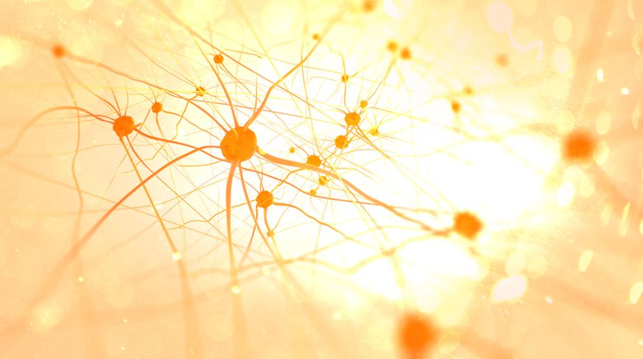
Representational image (Photo: Getty Images)
The internal volume of the cell exclusive of the nucleus is called the cytoplasm and is occupied by organelles and by the semifluid cytosol in which they are suspended.
Let’s take a look at each of the major eukaryotic organelles. In a typical animal cell, these compartments make up almost half of the total internal volume of the cell. As one continues on a tour of the eukaryotic cell and begins to explore its organelles, one may find it helpful to give these sub-cellular structures a human perspective and acquainting oneself with some of the hereditary human diseases that are associated with specific organelles.
Advertisement
In most cases, these disorders are caused by genetic defects in specific proteins — enzymes and transport proteins, most commonly — which are localised to particular organelles.
Advertisement
Our tour of the eukaryotic organelles begins with a very prominent organelle, the mitochondrion. Mitochondria are large by cellular standards — up to a micrometre across and usually a few micrometres long.
A mitochondrion is therefore comparable in size to a whole bacterial cell. The fact that most eukaryotic cells contain hundreds of mitochondria, each approximately the size of an entire bacterial cell, emphasises the great difference in size between prokaryotic and eukaryotic cells.
The mitochondrion is surrounded by two membranes, designated the inner and outer mitochondrial membranes. Most of the chemical reactions involved in the oxidation of sugars and other cellular “fuel” molecules occur within the mitochondria.
The purpose of these oxidative events is to extract energy from food and conserve as much of it as possible in the form of the high-energy compound Adenosine Tri- Phosphate. It is within the mitochondrion that the cell localises most of the enzymes and intermediates involved in such important cellular processes as the Tri-Carboxylic Acid cycle, fat oxidation, and ATP generation.
Most of the intermediates involved in the transport of electrons from oxidisable food molecules to oxygen are located in or on the cristae, in foldings of the inner mitochondrial membrane.
Other reaction sequences, particularly those of the TCA cycle and those involved in fat oxidation, occur in the semi-fluid matrix that fills the inside of the mitochondrion.
The number and location of mitochondria within a cell can often be related directly to their role in that cell. Tissues with an especially heavy demand for ATP as an energy source can be expected to have cells that are well endowed with mitochondria, and the organelles are usually located within the cell just where the energy need is greatest.
This localisation is illustrated by the sperm cell. As the drawing indicates, a sperm cell often has a single spiral mitochondrion wrapped around the central shaft, or axoneme, of the cell. Notice how tightly the mitochondrion coils around the axoneme, just where the ATP is actually needed to propel the sperm cell.
Muscle cells and cells that specialise in the transport of ions also have numerous mitochondria located strategically to meet the special energy needs of such cells. Fluorescent images of mitochondria in living cells show them to be ver y large and highlybranched. T
his type of structure is more widespread than just in sperm mid-pieces and may, in fact, be more representative of mitochondrial shape and size than most conventional diagrams indicate.
The writer is associate professor, head, department of botany, ananda mohan college, kolkata, and also fellow, botanical society of bengal, and can be contacted at tapanmaitra59@yahoo.co.in
Advertisement