The key disparities between prokaryotes and eukaryotes
Returning to the basic distinction between prokaryotes and eukaryotes, it recognises many important structural, biochemical, and genetic differences between eubacteria and archaea on the one hand and eukaryotic cells on the other.
Dr Tapan Kumar Maitra | New Delhi | June 3, 2024 1:43 pm
Prokaryotic cell (photo, SNS)
Returning to the basic distinction between prokaryotes and eukaryotes, it recognises many important structural, biochemical, and genetic differences between eubacteria and archaea on the one hand and eukaryotic cells on the other.
Presence or absence of a membrane-enveloped nucleus: The most fundamental distinction between eukaryotes and prokaryotes is reflected in the nomenclature itself. An eukaryotic cell has a true, membrane-bounded nucleus, whereas a prokaryotic cell does not. Instead of being enveloped by a membrane, the genetic information of a prokaryotic cell is localised in a region of the cytoplasm called the nucleoid. Within an eukaryotic cell, on the other hand, most of the genetic information is localised to the nucleus, which is surrounded not by a single membrane but by a nuclear envelope that consists of two membranes.
Use of internal membranes to segregate function: Prokaryotic cells contain few internal membranes; most cellular functions occur either in the cytoplasm or on the plasma (cell) membrane. By contrast, eukaryotic cells make extensive use of internal membranes to compartmentalise specific functions. Examples of internal membrane systems in eukaryotic cells include the endoplasmic reticulum, the Golgi complex, and the membranes that surround and delimit organelles such as mitochondria, chloroplasts, lysosomes, and peroxisomes, as well as various kinds of vacuoles and vesicles. Each of these organelles has its own characteristic membrane (or pair of membranes, in the case of mitochondria and chloroplasts), similar to other membranes in basic structure but often with its own distinctive chemical composition and proteins.
Advertisement
Tubules and filaments: Also found in the cytoplasm of eukaryotic cells are several non-membranous structures that are involved in cellular contraction and motility and in the establishment and support of cellular architecture. These include the microtubules found in the cilia and flagella of many cell types, the microfilaments of actin found in muscle fibrils and other structures involved in motility, and the intermediate filaments, which are especially prominent in cells that are subject to stress. Microtubules, microfilaments, and intermediate filaments are key components of the cytoskeleton that impart structure and elasticity to almost all eukaryotic cells.
Exocytosis and endocytosis: A further feature of eukaryotic cells is their ability to exchange materials between the membrane-bounded compartments within the cell and the exterior of the cell. This exchange is possible because of exocytosis and endocytosis, processes that are unique to eukaryotic cells. In endocytosis, portions of the plasma membrane invaginate and are pinched off to form membrane-bound cytoplasmic vesicles containing substances that were previously on the outside of the cell. Exocytosis is essentially the reverse of this process. Membrane-bounded vesicles inside the cell fuse with the plasma membrane and release their contents to the outside of the cell.
Organisation of DNA: Another distinction between prokaryotes and eukaryotes becomes apparent when considering the amount and organisation of the genetic material. Prokaryotes characteristically contain amounts of DNA that might be described as “reasonable”; that is, we can account for much of the DNA in terms of known proteins for which the DNA serves as a genetic blueprint. Prokaryotic DNA is usually present in the cell as one or more circular molecules with which relatively few proteins are associated. But if DNA appears to pose a packaging problem for prokaryotic cells, consider the case of the eukaryotic cell! Although some of the lower eukaryotes (such as yeasts and fruit flies) contain only 10 to 50 times as much DNA as bacteria, most eukaryotic cells have at least 1000 times as much DNA as E. coli. It is tempting to label such amounts of DNA as “unreasonable” because we cannot at present assign any known function to much of it. Whatever the genetic function of such large amounts of DNA, the packaging problem is clearly acute. It is solved universally among eukaryotes by the organisation of DNA into complex structures called chromosomes, which contain at least as much protein as DNA. It is as chromosomes that the DNA of eukaryotic cells is packaged, segregated during cell division, transmitted to daughter cells, and transcribed as needed into the molecules of RNA that are involved in protein synthesis.
Segregation of genetic information. A further contrast between prokaryotes and eukaryotes is the way they allocate genetic information to daughter cells upon division. Prokaryotic cells merely replicate their DNA and divide by a relatively simple process called cell fission, with one molecule of DNA going to each daughter cell. Eukaryotic cells also replicate their DNA, but they then use the more complex processes of mitosis and meiosis to distribute chromosomes equitably to daughter cells.
Expression of DNA. The differences between prokaryotic and eukaryotic cells extend to the expression of genetic information. Eukaryotic cells tend to transcribe genetic information in the nucleus into large RNA molecules and depend on later processing and transport processes to deliver RNA molecules of the proper sizes to the cytoplasm for protein synthesis. By contrast, prokaryotes transcribe very specific segments of genetic information into RNA messages, and little or no processing or selection appears to be either necessary or possible. In fact, the absence of a nuclear membrane makes it possible for new messenger RNA molecules to become involved in the process of protein synthesis even before they are themselves completely synthesised. Prokaryotes and eukaryotes also differ in the size and composition of the ribosomes used to synthesise proteins.
The author is an associate Professor (retd.) & former head of the Department of Botany at Ananda Mohan College.
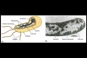
Structure of a typical bacterial cell: (a) A three-dimensional model showing the components of a typical bacterium, (b) An electron micrograph of a bacterial cell with several of the same components labeled. Notice that the nucleoid is simply a region within the cell, not a membrane-bounded compartment (TEM).
Ahead of the Parliament Session, Tamil Nadu Chief Minister and ruling DMK president MK Stalin has asked the party MPs to join hands with the INDIA bloc and take on the BJP-led Union Government to task.
During the event, senior advocates, advocates, and academicians will give a hands-on experience to the participating teams about real courtroom proceedings.
The accused, identified as Abhishek Kumar alias Harshu and Vikrant alias Vicky, were apprehended with two sophisticated pistols and 22 live rounds in their possession.